Tooth Decay Stages: 5 Stages, Signs, and What to Do

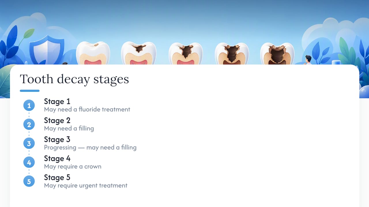
Tooth decay moves through five distinct stages: demineralization, enamel decay, dentin decay, pulp involvement, and abscess. The Cleveland Clinic confirms this five-stage framework, and where you fall on that spectrum determines everything about your treatment. The good news is that the first stage is reversible with fluoride and professional care. The bad news is that most people don’t notice anything until stage two or three, when a filling is already necessary.
If you have any of these symptoms right now, seek urgent dental care today:
Severe, throbbing tooth pain that doesn’t stop
Visible facial swelling or swelling in your jaw
Fever alongside dental pain
A bad taste in your mouth with pus or drainage
If none of those apply but you’ve noticed a white spot, mild sensitivity, or a dark area on a tooth, schedule a dental exam soon. Don’t wait for pain to appear. As Harvard Health notes, a large majority of adults have had at least one cavity, and early decay is often completely painless until it reaches the nerve.
Key Takeaways
Early-stage tooth decay is reversible with fluoride, but once a hole forms, every stage that follows requires progressively more complex dental treatment to resolve.
Point | Details |
Stage 1 is the only reversible stage | Demineralization (white spot lesion) can be stopped and reversed with fluoride before a hole forms. |
Pain is a late and unreliable sign | Decay can reach the dentin or even the pulp with no pain at all; routine X-rays catch what you can’t feel. |
Frequency of sugar exposure drives risk | Sipping sugary drinks slowly causes more acid attacks than consuming the same amount quickly at one sitting. |
Urgent signs require same-day care | Facial swelling, fever, or severe throbbing pain means seek dental or emergency care immediately. |
Mersaldental offers same-day emergency care | Fillings, crowns, root canals, and emergency appointments are available in downtown Ottawa with direct insurance billing. |
Table of Contents
How tooth decay actually starts
Tooth decay begins with bacteria. Your mouth naturally hosts hundreds of bacterial species, and certain ones, particularly Streptococcus mutans, feed on the sugars and starches you eat. When they do, they produce acid as a byproduct. That acid sits against your enamel and pulls out calcium and phosphate minerals in a process called demineralization. If it happens often enough and your saliva can’t keep up, a cavity forms.
The NIDCR explains this process clearly: bacteria in plaque use sugars to produce acids that attack enamel, and early demineralization can be stopped or reversed with fluoride. The key word is early. Once the enamel surface breaks down into a physical hole, the process is no longer reversible without a dentist.
Here’s the basic sequence:
Plaque forms on teeth within hours of eating, especially in grooves, between teeth, and near the gumline
Acid attacks begin each time you eat or drink something sugary or starchy
Minerals leach out of enamel during each acid attack, weakening the surface
Remineralization occurs between attacks when saliva deposits minerals back, but only if the attacks aren’t too frequent
Your saliva is your mouth’s natural defense. It neutralizes acid, delivers calcium and phosphate back to enamel, and contains antibacterial proteins. Fluoride from toothpaste and water strengthens enamel and makes it more resistant to acid. Regular professional cleanings remove the plaque your brush misses.
What raises your risk:
Sipping sugary drinks or snacking frequently throughout the day
Dry mouth from medications, medical conditions, or mouth breathing
Deep pits and fissures on back teeth that trap food
Infrequent brushing or skipping flossing
Acid reflux, which bathes teeth in stomach acid
Pit-and-fissure surfaces on molars are especially vulnerable. The grooves are narrow enough that toothbrush bristles can’t reach the bottom, so bacteria accumulate undisturbed. Smooth surfaces between teeth progress more slowly but are still at risk, particularly without flossing.
Pro Tip: Frequency of sugar exposure matters more than total quantity. Sipping one soda slowly over three hours causes more acid attacks than drinking it in ten minutes. Water between meals is one of the most practical ways to reduce your daily acid load.
The 5 stages of tooth decay: signs, symptoms, and treatment
Identifying which stage you’re in determines whether you need a fluoride treatment, a filling, a crown, or something more urgent. The five stages progress in one direction only, each one harder and more expensive to treat than the last.
Stage 1: Demineralization (white spot lesion)
What it looks like: A chalky white or off-white spot on the tooth surface, often near the gumline or in a groove. The enamel is still intact, but the mineral content underneath has dropped. Under good lighting, the spot may look dull compared to the surrounding tooth.
What it feels like: Usually nothing. Most people at this stage have no sensitivity, no pain, and no awareness that anything is wrong. Occasionally, a white spot near the gumline causes mild sensitivity to cold.
Treatment: This is the only stage that is fully reversible without drilling. Dentists apply fluoride varnish directly to the lesion, recommend prescription-strength fluoride toothpaste, and may suggest dietary changes. The NIDCR confirms that fluoride can stop and reverse early demineralization by helping enamel remineralize. Dental sealants on back teeth can also protect vulnerable grooves before lesions form.
What to do now: Book a dental exam. Mention the white spot specifically so the dentist can assess it and apply fluoride if appropriate.
Stage 2: Enamel decay
What it looks like: The white spot darkens to yellow or light brown as the enamel continues to break down. Eventually, the surface becomes rough or pitted. A small hole may be visible or felt with the tongue.
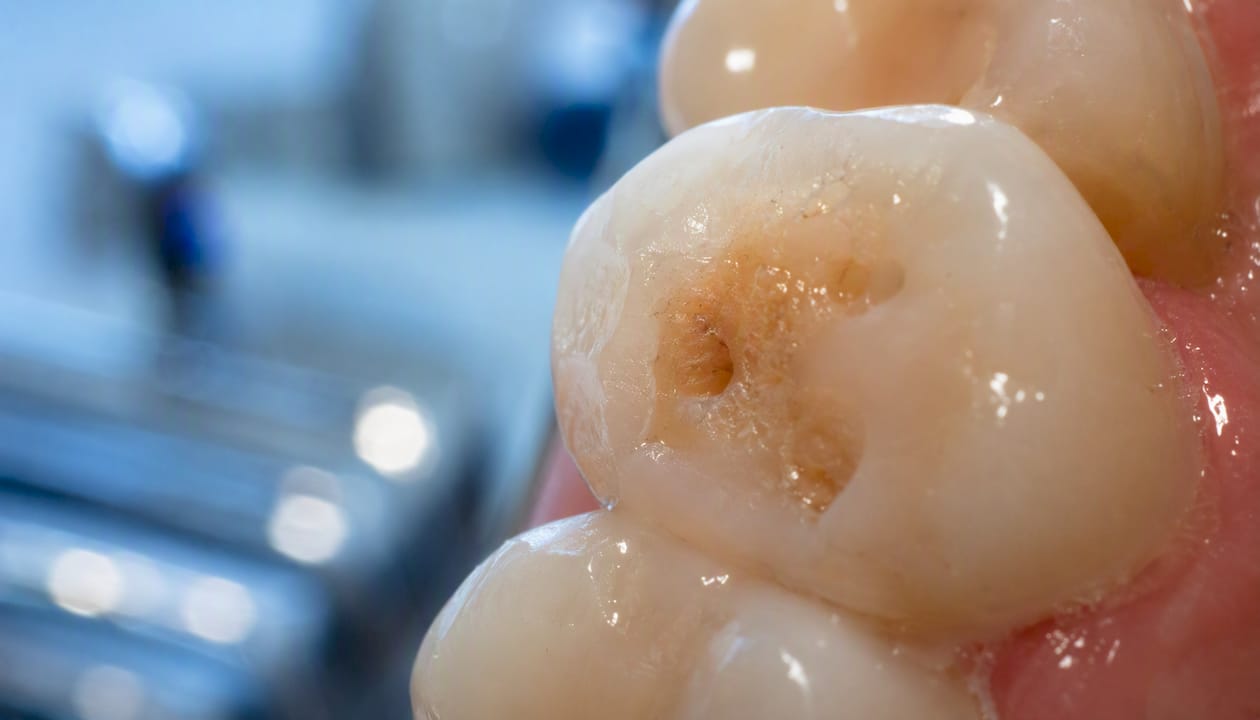
What it feels like: Mild sensitivity to sweets or cold is common. Some people feel a brief sharp sensation when eating something sugary. Many still feel nothing at all.
Treatment: Once the enamel surface has cavitated (broken through), remineralization alone won’t close the hole. A dentist removes the decayed material and places a filling. Composite resin (tooth-colored) is the standard choice for most visible teeth; amalgam is occasionally used in back teeth for durability. Mayo Clinic confirms that fillings are the appropriate intervention once a cavity has formed.
What to do now: Schedule a filling appointment. This is a routine procedure that takes one visit and stops the decay from advancing.
Stage 3: Dentin decay
What it looks like: A larger, darker cavity, often brown or black. The hole is more visible, and the surrounding tooth may look discolored. The decay has passed through enamel and entered dentin, the softer layer underneath.
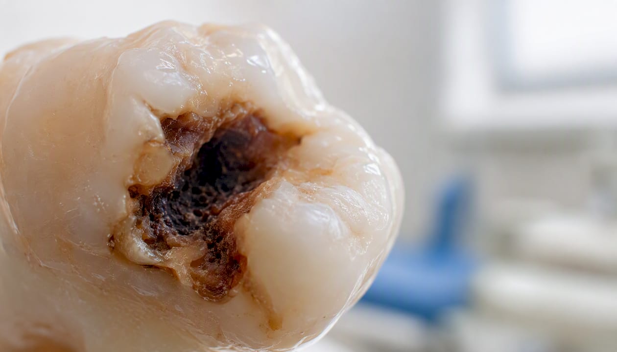
What it feels like: Noticeably more sensitivity, especially to cold, heat, and sweets. Sensitivity may linger for a few seconds after the trigger is removed. Some people experience a dull ache. If you’re noticing sudden sensitivity in one tooth, dentin involvement is a likely cause.
Treatment: A filling is still possible if enough tooth structure remains. Larger cavities may require a crown to protect the remaining tooth. The dentist removes all decayed dentin, shapes the cavity, and restores it. If sensitivity is significant, a desensitizing liner may be placed before the filling material. For more on managing sensitivity at this stage, treatment options for sensitive teeth cover what works clinically.
What to do now: See a dentist promptly. Dentin decay progresses faster than enamel decay, and waiting weeks can push the cavity closer to the pulp.
Stage 4: Pulp involvement
What it looks like: The cavity is large and often visibly dark. The tooth may look fractured or significantly broken down. At this point, the decay has reached the pulp chamber, where the nerve and blood vessels live.
What it feels like: Spontaneous pain that doesn’t need a trigger. Throbbing, persistent aching, or sharp pain when biting. The tooth may be extremely sensitive to heat, and cold may actually provide brief relief (a classic sign of irreversible pulpitis). Some people experience numbness or pressure when biting down, which can signal nerve involvement.
Treatment: A root canal is the standard treatment to save the tooth. The dentist removes the infected pulp tissue, cleans and shapes the root canals, fills them with a biocompatible material, and places a crown to protect the remaining structure. Signs that a root canal is needed include spontaneous pain, prolonged sensitivity to heat, and discoloration. The root canal treatment page explains what the procedure involves and what to expect.
What to do now: Call your dentist as soon as possible. Pulp involvement is not an emergency if there’s no swelling or fever, but it will not improve on its own and will progress to an abscess without treatment.
Stage 5: Abscess
What it looks like: Severe swelling of the gum, jaw, or face. A visible pimple-like bump on the gum (called a fistula or sinus tract) may be present, sometimes draining pus. The tooth may look dark or fractured.
What it feels like: Intense, throbbing pain that may radiate to the jaw, ear, or neck. Fever, swollen lymph nodes, difficulty swallowing, and a foul taste in the mouth. Some people feel a sudden relief of pressure if the abscess drains on its own, but the infection is still present.
Treatment: Urgent dental care is required. The American Dental Association’s MouthHealthy resource describes abscess formation as a pocket of pus at the root tip that can cause facial swelling and systemic signs, requiring prompt intervention. Treatment options include draining the abscess, root canal therapy to save the tooth, or extraction if the tooth cannot be restored. Antibiotics may be prescribed when infection has spread, but they do not replace dental treatment. If facial swelling is spreading or you have difficulty breathing or swallowing, go to an emergency room immediately.
What to do now: Seek same-day dental care or go to an emergency room if swelling is spreading rapidly.
Pro Tip: A cavity at stage 2 or 3 often causes no pain at all. Waiting until something hurts before seeing a dentist is the single most common reason a simple filling becomes a root canal.
How you and a dentist determine which stage you have
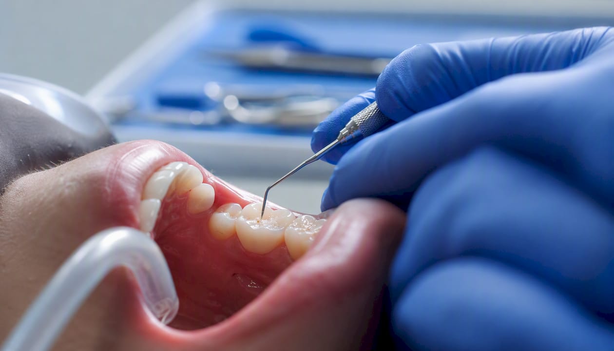
Some signs can be spotted at home, but confirming how deep decay has progressed requires a professional exam and, in most cases, X-rays. A white spot you can see in the mirror might be stage 1. A dark spot that feels rough to your tongue is likely stage 2 or 3. Pain without a trigger points to stage 4. Swelling means stage 5.
Home signs worth paying attention to:
White, chalky spots on any tooth surface (stage 1)
Dark spots, brown or black discoloration (stage 2–3)
Rough or pitted texture you can feel with your tongue
Sensitivity to cold, heat, or sweets that lingers
A visible hole or broken tooth surface
Spontaneous pain or throbbing without eating or drinking
Swelling in the gum, jaw, or face
A bad taste or pus near a tooth
At the dental office, the dentist combines a visual exam, a tactile probe of the tooth surface, and radiographs to confirm the stage. Bitewing and periapical X-rays are the key diagnostic tools for this: bitewings reveal decay between teeth and the depth of enamel and dentin involvement, while periapical films show the root tip and surrounding bone, which is where abscesses appear. Some interproximal cavities (between teeth) are completely invisible without X-rays, even to an experienced clinician.
Pro Tip: Pain is a late and unreliable indicator of decay severity. Decay can destroy most of the dentin and reach the pulp while causing no discomfort at all. Routine X-rays every 12–24 months catch lesions that neither you nor your dentist can see on visual exam alone.
How fast decay progresses and what speeds it up
Decay doesn’t move at a fixed rate. Enamel penetration can take two to three years in a low-risk patient with good saliva flow and regular fluoride exposure. Once decay reaches dentin, progression accelerates significantly, and dentin-to-pulp involvement can happen in as little as a year. MSD Manuals notes that pit-and-fissure decay on back teeth often progresses faster than smooth-surface decay because grooves trap bacteria and are harder to clean.
Factors that speed up decay:
Frequent sugar or starch exposure throughout the day
Low saliva flow (dry mouth from medications, radiation therapy, or systemic disease)
Deep pits and fissures on molars and premolars
Infrequent brushing and no flossing
Acid reflux or frequent vomiting (eating disorders)
Previous cavities (higher baseline bacterial load)
Factors that slow decay:
Consistent fluoride exposure from toothpaste, water, and professional varnish
Good salivary flow, which neutralizes acid and remineralizes enamel
Dental sealants on back teeth
Professional cleanings every six months to remove plaque buildup
Limiting sugar frequency rather than total intake
Prevalence note: Harvard Health reports that roughly 90% of adults have had at least one cavity, making dental caries one of the most common chronic conditions worldwide. Early decay is often asymptomatic, which is why routine exams catch what patients miss.
The practical takeaway: a stage 1 lesion in a low-risk patient with good hygiene may stay stable for years. The same lesion in someone with dry mouth, frequent snacking, and no professional care can reach the pulp within 12–18 months.
Quick reference: treatment by stage and urgency
Match your signs to the stage below to understand what treatment typically involves and how quickly you should act.
Stage | Common treatment | What you may notice | Urgency |
Stage 1: Demineralization | Fluoride varnish, dietary counseling, prescription fluoride toothpaste | White or chalky spot, no pain | Routine — book within weeks |
Stage 2: Enamel decay | Composite or amalgam filling (one visit) | Mild sensitivity, small visible hole or dark spot | Routine — book within 1–2 weeks |
Stage 3: Dentin decay | Filling or crown depending on size | Sensitivity to cold/heat/sweets, larger dark cavity | Soon — book within days to 1 week |
Stage 4: Pulp involvement | Root canal + crown | Spontaneous pain, throbbing, heat sensitivity | Prompt — call same day or next day |
Stage 5: Abscess | Drain abscess + root canal or extraction, possible antibiotics | Swelling, fever, severe pain, pus | Urgent — same-day care required |
On material choices: composite resin is the standard for most fillings today because it bonds directly to tooth structure and matches tooth color. Amalgam remains an option for large cavities in back teeth where durability is the priority. When decay has destroyed enough tooth structure that a filling would leave the remaining walls at risk of fracture, a crown is the appropriate restoration. Crowns are also placed after root canals on back teeth to protect the now-brittle tooth from cracking under chewing forces.
When in doubt about which stage applies to you, a dental exam is the only reliable answer. Rapid progression cases, especially in patients with dry mouth or high sugar intake, warrant prompt assessment rather than a wait-and-see approach.
How to stop or reverse early decay and reduce future risk
The first stage of tooth decay is the only one you can reverse without a drill. Demineralization responds to fluoride because fluoride incorporates into the enamel crystal structure and makes it more resistant to future acid attacks. Once a hole forms, that window closes.
Daily habits that protect your teeth:
Brush twice daily with a fluoride toothpaste, spending at least two minutes each session
Floss once daily to remove plaque from between teeth where your brush can’t reach
Reduce the frequency of sugary or starchy snacks and drinks, not just the total amount
Drink water after meals to rinse away food and acid
Chew sugar-free gum containing xylitol after meals to stimulate saliva flow
Professional prevention options:
Fluoride varnish: applied at hygiene appointments, it delivers a concentrated dose directly to enamel and is especially effective for early lesions
Dental sealants: the NIDCR recommends sealants for the grooves and pits of back teeth, where toothbrush bristles can’t reach; they’re most effective when applied soon after permanent molars erupt
Prescription fluoride gels or rinses: for high-risk patients, including those with dry mouth or a history of frequent cavities
Regular hygiene appointments: professional cleaning removes calcified plaque (tartar) that brushing can’t touch, and the hygienist can catch early lesions before they progress
Booking a hygiene appointment is the single most effective professional step for prevention. The hygienist removes what you can’t, applies fluoride, and gives the dentist a chance to catch stage 1 lesions before they need a filling.
Pro Tip: Timing your sugar exposure matters more than most people realize. A glass of orange juice with breakfast causes one acid attack. Sipping it slowly over an hour causes a sustained attack that overwhelms saliva’s ability to remineralize. Eat sweets with meals rather than between them, and rinse with water immediately after.
When to schedule a routine visit versus urgent care
Seek urgent dental care the same day if you have severe pain, swelling in your face or jaw, fever, or a bad taste with drainage. For everything else, book a standard appointment within days to a couple of weeks depending on your symptoms.
Go to the dentist today or to an emergency room if you have:
Facial or jaw swelling that is spreading
Fever with dental pain
Difficulty swallowing or breathing
Severe, uncontrolled throbbing pain
Pus or drainage from around a tooth
Book a standard dental appointment within 1–2 weeks if you notice:
A white or chalky spot on a tooth
A dark spot, rough area, or visible hole
Sensitivity to cold, heat, or sweets that wasn’t there before
A tooth that feels different when you bite down
Persistent bad breath that doesn’t improve with brushing
At the appointment, the dentist will do a visual and tactile exam, take X-rays if needed, review your symptoms and dental history, and present a treatment plan. For urgent visits, pain control (local anesthetic, temporary sedation, or a prescription) is addressed first, then the definitive treatment is either completed the same day or scheduled promptly. Emergency dental care is available for patients who need same-day attention.
Complications of untreated tooth decay
Untreated decay doesn’t stabilize. It progresses to pulp infection, abscess, bone loss, and eventual tooth loss. In rare cases, a dental abscess can spread to the jaw, neck, or airway, which is a life-threatening emergency.
What can happen when decay goes untreated:
Pulp infection: bacteria reach the nerve and blood supply, causing irreversible inflammation and death of the pulp tissue
Dental abscess: a pocket of pus forms at the root tip, causing severe pain, swelling, and potential spread to surrounding structures
Tooth fracture: extensive decay weakens the tooth structure until it cracks or breaks, often making restoration more complex or impossible
Spread to adjacent teeth: bacterial load increases and neighboring teeth become more vulnerable
Jawbone involvement: infection can spread into the surrounding bone (osteomyelitis), requiring more aggressive treatment
Systemic infection: in rare but serious cases, oral bacteria enter the bloodstream and can cause sepsis
The cost and complexity of treatment rises sharply with each stage. A stage 2 filling costs a fraction of a stage 4 root canal and crown. A stage 5 abscess requiring extraction, followed by an implant or bridge to replace the tooth, is the most expensive outcome of all. Dental implants and dentures are the main replacement options after tooth loss, but neither is as good as keeping the natural tooth.
How our dentists assess and choose treatments
At Mersaldental, the diagnostic process combines three things: a visual and tactile exam, radiographs, and a conversation about your symptoms and history. No single tool tells the whole story. A tooth that looks fine on the surface can have significant interproximal decay visible only on a bitewing X-ray. A tooth that looks dark may be stained rather than decayed. The clinical picture only becomes clear when all three sources of information are combined.
What a typical visit looks like:
A hygiene visit often catches early lesions before you’ve noticed any symptoms
The dentist probes the tooth surface for softness, checks for cavitation, and reviews X-rays
Treatment is staged by urgency: acute pain and abscesses are addressed first; restorative work (fillings, crowns) is scheduled based on severity and patient preference
Same-day fillings are available when the cavity is straightforward and the schedule allows
Root canals and crowns are scheduled as dedicated appointments
Questions worth bringing to your appointment:
Is this lesion at a stage where fluoride treatment could still reverse it?
Do I need a filling now, or can we monitor this for a few months?
What material do you recommend for this filling and why?
If I need a crown, what does the process involve?
Is this tooth saveable, or is extraction the better long-term option?
At Mersaldental, we use clinical staging, not just appearance, to decide between remineralization and operative treatment. A patient with high caries risk may receive a restoration for a lesion that looks shallow, because the risk of rapid progression outweighs the benefit of waiting. The goal is always to conserve as much natural tooth structure as possible.
Mersaldental accepts direct insurance billing, the Canadian Dental Care Plan (CDCP), and the Interim Federal Health Program (IFHP), so cost is rarely a reason to delay care. Same-day emergency appointments are available for acute pain and swelling.
What we emphasize when treating decay
Prevention and early detection are where the real difference is made. By the time a tooth hurts, the treatment is almost always more involved than it needed to be. The patients who do best are the ones who come in for routine hygiene appointments, mention new sensitivity early, and don’t wait for a crisis.
When decay is caught at stage 1 or 2, outcomes are predictable and treatment is straightforward. A fluoride application or a simple filling, done well, can protect a tooth for years. That’s the outcome we work toward for every patient, and it’s entirely achievable with regular care.
Dental care for tooth decay at Mersaldental
If you suspect advanced decay or you’re experiencing pain, sensitivity, or swelling, the right move is to get it assessed before it progresses further. At Mersaldental in downtown Ottawa, we offer same-day emergency appointments for acute dental pain and swelling, along with the full range of treatments for every stage of decay.

Our services for decay-related care include fillings, crowns and bridges, root canal treatment, extractions, and preventive hygiene appointments with fluoride varnish. We accept direct insurance billing, the Canadian Dental Care Plan (CDCP), and the Interim Federal Health Program (IFHP), so you can focus on your care rather than the paperwork. New patients are welcome.
Book an appointment or learn more about our services — or call us directly if you need same-day care. The earlier we see you, the more options we have.
Frequently asked questions about tooth decay stages
Can a cavity heal on its own? Stage 1 demineralization can reverse with fluoride and reduced acid exposure. Once a physical hole forms in the enamel (stage 2 or beyond), the cavity cannot heal on its own and requires a dental restoration.
How do I know if I have a cavity without going to the dentist? You may notice a white or dark spot, a rough area, sensitivity to cold or sweets, or a visible hole. But many cavities, especially between teeth, are invisible and painless until they’re large. A dental exam with X-rays is the only reliable way to confirm.
How long does it take for a cavity to go from enamel to the nerve? It varies. Enamel penetration can take two to three years in a low-risk patient. Dentin-to-pulp progression can happen in as little as a year, particularly in patients with dry mouth, frequent sugar exposure, or poor oral hygiene.
What does a stage 1 cavity look like? A chalky white or off-white spot on the tooth surface, often near the gumline or in a groove. The enamel is still intact, but the area looks dull compared to the surrounding tooth.
Is a root canal always needed for a deep cavity? Not always. If the decay has reached the pulp, a root canal is typically necessary to save the tooth. If the cavity is deep but hasn’t reached the pulp, a filling or crown may be sufficient, sometimes with a protective liner placed near the pulp.
What happens if a tooth abscess goes untreated? The infection can spread to the jaw, neck, or airway. In rare cases, it becomes a life-threatening emergency. Facial swelling that is spreading, fever, or difficulty swallowing requires immediate emergency care.
Does the CDCP cover cavity treatment? The Canadian Dental Care Plan covers a range of dental services for eligible Canadians. Mersaldental accepts CDCP and can help you understand what your coverage includes at your appointment.
This article is general information, not a substitute for advice from a qualified doctor. Consult a qualified healthcare professional about your own circumstances before acting on anything here.
Sources
These are the reputable clinical and public health sources used to build this article. Each one offers reliable, evidence-based information on tooth decay, diagnosis, and treatment.
Recommended
Comments